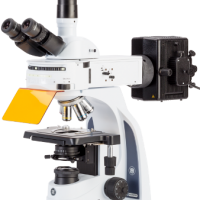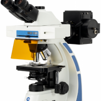
For biological and medical applications, microscopes equipped for fluorescence are used to observe certain parts of living cells and tissues with help of fluorophores.
One or more fluophores are added to certain parts of the specimen tissue or cells. When exposed to so-called “exicitation light”, the fluorophore absorbs energy from the excitation light and emits ligth in another visible wavelengt. This make it possible to distinguish tumor cells from other cells, to prove the presence or absence of antibodies or to observe sub-microscopic structures of cells and tissues,etc
Euromex fluorescence microscopes are constructed with an incident light source, specific Plan or Plan Fluarex – optional semi-apochromatic – objectives, a set of excitation and emission filters and a dichroic mirror
– The upright models have a modern 3 Watt LED transmitted light source and a 100 W incident mercury vapor illuminator.
– The inverted models have a 100 W transmitted mercury vapor illuminator and a 30 W halogen incident light source.